previous image | main article | next image
CT Scans in Art Work Appraisal
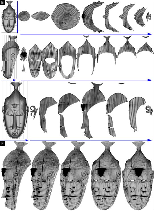
Baule mask, Ivory
Coast. Wood, 29 cm. E. CT: thin sections of the same Baule mask as in the previous figure, taken on the three spatial planes. The growth rings in the wood are clearly visible, as are the nail tips and metal triangles on the forehead which have caused distortion in the images due to the diffraction of the X-rays on the crystalline structure of the metal. F. CT: translucent 3D views taken while the mask was rotated around a vertical axis, bringing out the grain of the wood. Back: Figure 1 |
previous image | main article | next image